-
Use Cases
-
Resources
-
Pricing
The history of microscopy traces the development of instruments used to see objects too small for the naked eye. Beginning with simple magnifying glasses in ancient times and the compound microscope of the late 16th century, the field advanced dramatically through the work of pioneers like Robert Hooke and Antonie van Leeuwenhoek. The 19th century brought improved optics and cell theory, while the 20th century introduced electron microscopes capable of atomic-scale resolution, and modern techniques like fluorescence and super-resolution microscopy continue to revolutionize biology and medicine. More Less
1286
% complete
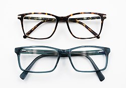
Eyeglasses for correcting vision appeared in northern Italy around this time, marking the beginning of practical lens grinding and polishing techniques that would eventually enable magnifying instruments.
Image source: Glasses
65
% complete
The Roman philosopher Seneca noted that small letters could be seen larger through a glass globe filled with water, an early recorded observation of magnification by refraction that foreshadowed the lens.
Image source: History of optics
1590
% complete
Dutch spectacle-maker Zacharias Janssen is often credited with creating one of the earliest compound microscopes, using multiple lenses in a tube to achieve greater magnification than a single lens could provide.
Image source: Zacharias Janssen
1609
% complete
Galileo adapted his telescope design into a compound microscope, which he called the 'occhiolino' or 'little eye', demonstrating that optical instruments could be used to examine tiny objects as well as distant ones.
Image source: Galileo Galilei
1625
% complete
Giovanni Faber, a member of the Accademia dei Lincei, coined the word 'microscope' from the Greek words for 'small' and 'to look at', giving the instrument its enduring name.
Image source: Microscopy
1661
% complete
Italian physician Marcello Malpighi used the microscope to discover capillaries connecting arteries and veins, providing crucial evidence for William Harvey's theory of blood circulation.
Image source: Marcello Malpighi
1665
% complete
Robert Hooke's Micrographia showcased stunning drawings of microscopic objects and coined the term 'cell' after observing box-like structures in cork, inspiring generations of microscopists.
Image source: Micrographia
1674
% complete
Using his handcrafted single-lens microscopes capable of over 200x magnification, van Leeuwenhoek observed 'animalcules' — bacteria, protozoa, and other microorganisms — becoming the father of microbiology.
Image source: Antonie van Leeuwenhoek
Oct 9, 1676
% complete
Van Leeuwenhoek's letter describing 'little animals' in water was presented to the Royal Society in London, initially met with skepticism but ultimately verified by Hooke and others, revolutionizing biology.
Image source: Royal Society
1700
% complete
London instrument maker John Marshall introduced improved focusing mechanisms and more stable stands, making compound microscopes easier to use and helping establish microscopy as a serious scientific tool.
1744
% complete
London maker John Cuff designed a practical, stable microscope with fine focus controls that became widely copied, standardizing microscope design across Europe in the mid-18th century.
1768
% complete
Spallanzani used microscopic observation alongside boiling experiments to challenge the theory of spontaneous generation, advancing the use of microscopy in experimental biology.
Image source: Lazzaro Spallanzani
1820
% complete
Building on work by Charles Chevalier and others, achromatic objective lenses combining crown and flint glass dramatically reduced color distortion, greatly improving image clarity in compound microscopes.
Image source: Achromatic lens
1827
% complete
Botanist Robert Brown observed the erratic movement of pollen grains suspended in water under his microscope, a phenomenon later explained by Einstein as molecular motion.
Image source: Brownian motion
1830
% complete
Lister published calculations showing how properly spaced achromatic lenses could eliminate spherical aberration, enabling high-quality compound microscopes and transforming biological research.
Image source: Joseph Jackson Lister
1838 - 1839
% complete
Using improved microscopes, Schleiden and Schwann proposed that all living things are composed of cells, establishing microscopy as central to biology and founding modern cytology.
Image source: Cell theory
1866
% complete
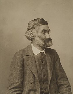
Physicist Ernst Abbe partnered with optician Carl Zeiss in Jena, Germany, applying rigorous wave-optics theory to lens design and launching a new era of scientifically engineered microscopes.
Image source: Ernst Abbe
1873
% complete
Abbe derived the theoretical limit of optical resolution based on diffraction and numerical aperture, defining the fundamental constraints of light microscopy for over a century.
Image source: Diffraction-limited system
1877
% complete
The Abbe condenser concentrated and controlled light passing through specimens, dramatically improving image brightness and contrast and becoming a standard component of laboratory microscopes.
Image source: Condenser (optics)
1882
% complete
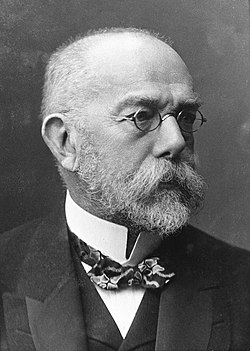
Koch combined oil immersion microscopy with staining techniques to visualize Mycobacterium tuberculosis, demonstrating microscopy's power in medical microbiology and germ theory.
Image source: Robert Koch
1893
% complete
August Köhler devised an illumination method providing even, controllable lighting of specimens, which remains the standard technique for optimal image quality in transmitted-light microscopy.
Image source: Köhler illumination
1932
% complete
Frits Zernike's phase-contrast technique converted phase shifts into visible brightness differences, allowing transparent living cells to be seen without staining; he won the Nobel Prize in Physics in 1953.
Image source: Phase-contrast microscopy
1955
% complete
Nomarski's DIC microscopy created pseudo-three-dimensional images of transparent specimens with striking relief, becoming invaluable for observing unstained living cells and fine structures.
Image source: Differential interference contrast microscopy
1931
% complete
At the Technical University of Berlin, Knoll and Ruska demonstrated that magnetic coils could focus electron beams to form magnified images, breaking the diffraction barrier of light microscopes.
Image source: Electron microscope
1937
% complete
Von Ardenne developed the first scanning electron microscope, rastering a focused electron beam across samples; SEM technology matured commercially in the 1960s to produce detailed surface images.
Image source: Scanning electron microscope
1938
% complete
Siemens produced the first commercial transmission electron microscope (TEM), achieving resolutions far beyond light optics and opening the ultrastructure of cells, viruses, and materials to direct observation.
Image source: Transmission electron microscopy
1981
% complete
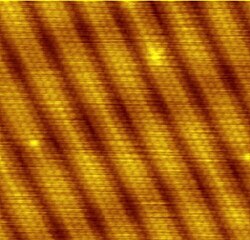
At IBM Zurich, Binnig and Rohrer created the STM, imaging surfaces atom by atom via quantum tunneling current. They shared the 1986 Nobel Prize in Physics for this breakthrough in nanoscale imaging.
Image source: Scanning tunneling microscope
1986
% complete
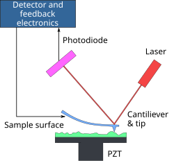
The atomic force microscope used a sharp tip on a flexible cantilever to feel surface topography with atomic resolution, extending probe microscopy to non-conductive materials including biological samples.
Image source: Atomic force microscopy
1994
% complete
Martin Chalfie demonstrated GFP as a genetic fluorescent tag in living organisms, revolutionizing live-cell imaging; Chalfie, Osamu Shimomura, and Roger Tsien shared the 2008 Nobel Prize in Chemistry.
Image source: Green fluorescent protein
2000
% complete
Stefan Hell's STED technique bypassed the diffraction limit using patterned light to switch fluorophores, achieving nanometer resolution and launching the super-resolution microscopy revolution.
Image source: STED microscopy
2006
% complete
Eric Betzig's PALM and William Moerner's foundational single-molecule spectroscopy enabled localization-based super-resolution imaging; they shared the 2014 Nobel Prize in Chemistry with Stefan Hell.
Image source: Photoactivated localization microscopy
2013
% complete
Advances in direct electron detectors and computational processing made cryo-EM capable of near-atomic-resolution structures of proteins, transforming structural biology; Dubochet, Frank, and Henderson won the 2017 Nobel Prize in Chemistry.
Image source: Cryo-electron microscopy
Or browse the full history timeline directory, with more than 2,000 topics.
This History of Microscopy timeline was generated with the help of AI, using information found on the internet.
We work hard to keep these timelines accurate, but mistakes do get through. If you spot one, email us at [email protected] and we'll fix it for future visitors.
Generate yours with AI or build it from scratch, for free.
Export your timeline, add your own events, edit or remove AI-generated events, and much more
No credit card required.
Cancel anytime.
Cancel anytime.
You can create an unlimited number of timelines with up to 10 events on each one, customize how they look, export them to PDF, PNG, PowerPoint, CSV, and Excel, and share them with a link. Paid plans raise the event limit and add features like spreadsheet imports and collaborators.
Yes. You can cancel your subscription from your account page at any time, and you will not be charged again. Your subscription stays active for the rest of the period you already paid for.
Yes. We will email you a reminder before the annual renewal, and we will also email you a receipt.
Yes. You can email us within 15 days of any payment, and we will issue you a full refund.
Check out our pricing docs or send us an email anytime: [email protected].