-
Use Cases
-
Resources
-
Pricing
The history of radiology traces the development of medical imaging from Wilhelm Conrad Röntgen's accidental discovery of X-rays in 1895, through the early use of fluoroscopy and contrast agents, to revolutionary technologies such as computed tomography, magnetic resonance imaging, ultrasound, and digital radiography that transformed diagnosis and treatment. More Less
Nov 8, 1895
% complete
German physicist Wilhelm Röntgen discovered X-rays while experimenting with cathode ray tubes, observing a mysterious radiation that could pass through matter and expose photographic plates. He named the rays 'X' for their unknown nature and was awarded the first Nobel Prize in Physics in 1901 for this groundbreaking discovery.
Image source: X-ray
Dec 22, 1895
% complete
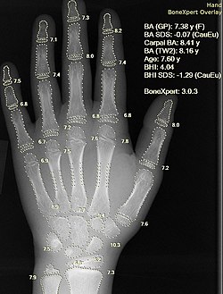
Röntgen produced the first radiograph, an image of his wife Anna Bertha's hand showing her bones and wedding ring. The striking image demonstrated the medical potential of X-rays and is often considered the birth of diagnostic imaging.
Image source: Wilhelm Röntgen
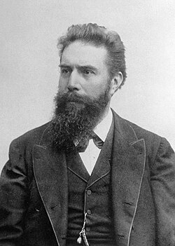
Feb 1896
% complete
Within weeks of Röntgen's announcement, surgeons in Germany and the United States began using X-rays to locate foreign objects such as bullets and needles in patients, marking the first practical medical applications of radiography.
Image source: Radiology
Mar 1896
% complete
French physicist Henri Becquerel discovered that uranium salts emitted penetrating radiation without any external energy source. This discovery of natural radioactivity complemented Röntgen's work and opened new avenues in medical physics.
Image source: Henri Becquerel
Nov 1896
% complete
During the First Italo-Ethiopian War, Italian military physicians used X-rays to locate bullets and shrapnel in wounded soldiers, demonstrating the value of radiography on the battlefield and foreshadowing its widespread wartime adoption.
Image source: Radiography
Dec 21, 1898
% complete
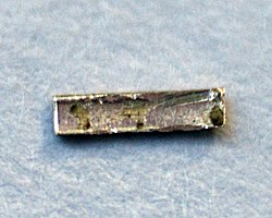
The Curies isolated radium from pitchblende ore, discovering one of the most intensely radioactive elements known at the time. Their work laid the foundation for radiotherapy and earned them the Nobel Prize in Physics in 1903 alongside Becquerel.
Image source: Radium
1899
% complete
Swedish physician Tor Stenbeck and others successfully used X-rays to treat skin cancer, demonstrating that radiation could destroy malignant tissue. This pioneering treatment established the field of radiation oncology.
Image source: Radiation therapy
1900
% complete
The American Roentgen Ray Society (ARRS) was established as the first radiological society in North America, providing a professional organization for early practitioners of the emerging specialty and publishing the American Journal of Roentgenology.
1898
% complete
Researchers including Walter Cannon pioneered the use of bismuth and later barium sulfate as oral contrast media, allowing visualization of the stomach and intestines and transforming the diagnosis of gastrointestinal diseases.
Image source: Barium sulfate suspension
1927
% complete
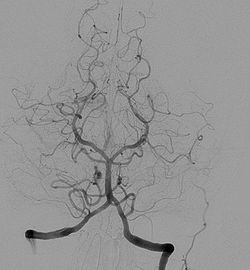
Portuguese neurologist António Egas Moniz performed the first cerebral angiograms by injecting contrast dye into blood vessels to visualize brain circulation. His technique enabled the diagnosis of tumors and vascular abnormalities and won him recognition beyond his controversial lobotomy work.
Image source: Cerebral angiography
1928
% complete
Greek-American physician Georgios Papanikolaou presented his findings on detecting cervical cancer through microscopic examination of vaginal smears, establishing cytology-based cancer screening that became standard preventive care.
Image source: Pap test
1929
% complete
German surgeon Werner Forssmann inserted a catheter into his own heart via his arm vein and confirmed its position with an X-ray image. His audacious self-experiment laid the groundwork for modern interventional cardiology and earned him the Nobel Prize in 1956.
Image source: Cardiac catheterization
1930
% complete
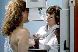
German radiologist Albert Salomon published studies using X-rays of breast specimens to detect breast cancer, initiating the development of mammography as a dedicated imaging technique that would later become central to breast cancer screening.
Image source: Mammography
1958
% complete
Cardiologist F. Mason Sones accidentally injected contrast directly into a patient's coronary artery during a cardiac catheterization at the Cleveland Clinic and found it was tolerated safely. This accident led to selective coronary angiography, revolutionizing heart disease diagnosis.
Image source: Coronary CT angiography
1963
% complete
Charles Dotter performed the first percutaneous transluminal angioplasty in Portland, Oregon, dilating a blocked leg artery without open surgery. His minimally invasive approach gave rise to interventional radiology, encompassing stenting, embolization, biopsy, and drainage procedures guided by imaging.
Image source: Interventional radiology
1976
% complete
Large randomized trials beginning with the HIP study demonstrated that population-based mammographic screening reduced breast cancer mortality, leading governments worldwide to establish organized screening programs for women.
1958
% complete
Scottish physician Ian Donald published pioneering work applying ultrasound to visualize the fetus in utero. Sonography quickly became indispensable for monitoring pregnancy, assessing fetal development, and diagnosing complications without ionizing radiation.
Image source: Obstetric ultrasonography
1958
% complete
Hal Anger invented the gamma camera, allowing functional imaging of organs after injection of radioactive tracers such as technetium-99m. Nuclear medicine enabled assessment of organ function rather than just anatomy.
Image source: Gamma camera
1971
% complete
British engineer Godfrey Hounsfield built the first computed tomography scanner at EMI, producing cross-sectional images of the head. Combined with Allan Cormack's mathematical contributions, CT transformed medicine and earned both inventors the 1979 Nobel Prize in Physiology or Medicine.
Image source: CT scan
Oct 1, 1971
% complete
At Atkinson Morley Hospital in London, the first whole-body clinical CT examination was performed on a patient with a suspected frontal lobe tumor, revealing a cyst clearly. The success of this scan heralded a new era of non-invasive diagnostic imaging.
Image source: History of computed tomography
1974
% complete
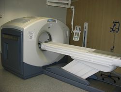
Michael Phelps and colleagues at Washington University developed the first positron emission tomography scanner. PET allowed visualization of metabolic activity using radiotracers like fluorodeoxyglucose, becoming vital in oncology, neurology, and cardiology.
Image source: Positron emission tomography
1977
% complete
Raymond Damadian and colleagues obtained the first full-body MRI scan of a human, taking nearly five hours to acquire. Building on Paul Lauterbur and Peter Mansfield's foundational work, MRI provided unprecedented soft-tissue contrast without ionizing radiation; Lauterbur and Mansfield shared the 2003 Nobel Prize in Physiology or Medicine.
Image source: Magnetic resonance imaging
1980 - 1999
% complete
Computed radiography and later direct digital radiography systems replaced analog film, enabling instant image acquisition, post-processing, electronic storage, and transmission. This transition dramatically improved workflow and image accessibility across healthcare systems.
1982
% complete
Picture archiving and communication systems (PACS) were deployed to store, retrieve, and distribute digital medical images across hospital networks, eliminating physical film archives and enabling remote interpretation by radiologists.
Image source: Picture archiving and communication system
1989
% complete
Kalender and colleagues introduced spiral CT, in which the table moves continuously while the tube rotates, allowing rapid volumetric acquisition of entire organs within a single breath-hold and enabling three-dimensional reconstruction.
1991
% complete
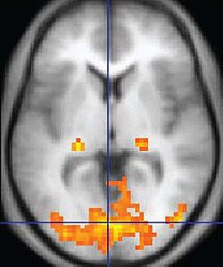
Researchers used BOLD contrast to visualize changes in brain blood oxygenation associated with neural activity, creating fMRI. This technique revolutionized cognitive neuroscience by allowing non-invasive mapping of human brain function.
Image source: Functional magnetic resonance imaging
1998
% complete
Four-slice multidetector CT scanners entered clinical use, dramatically reducing scan times. Subsequent advances led to non-invasive coronary CT angiography and whole-body trauma imaging, expanding CT's role across medicine.
Feb 2011
% complete
The FDA approved the first digital breast tomosynthesis system, which acquires multiple low-dose images reconstructed into thin slices of the breast. Tomosynthesis reduces false positives and improves cancer detection compared to conventional two-dimensional mammography.
Image source: Tomosynthesis
2017
% complete
Convolutional neural networks trained on large imaging datasets achieved expert-level performance detecting lung nodules, diabetic retinopathy, and other conditions, ushering artificial intelligence into routine radiology workflows as decision-support tools.
Image source: Artificial intelligence in healthcare
Sep 2021
% complete
The FDA cleared the first photon-counting detector CT system for clinical use. By counting individual X-ray photons and measuring their energy, this technology offers higher spatial resolution, lower radiation dose, and improved spectral imaging capabilities over conventional detectors.
Image source: Photon-counting computed tomography
Or browse the full history timeline directory, with more than 2,000 topics.
This History of Radiology timeline was generated with the help of AI, using information found on the internet.
We work hard to keep these timelines accurate, but mistakes do get through. If you spot one, email us at [email protected] and we'll fix it for future visitors.
Generate yours with AI or build it from scratch, for free.
Export your timeline, add your own events, edit or remove AI-generated events, and much more
No credit card required.
Cancel anytime.
Cancel anytime.
You can create an unlimited number of timelines with up to 10 events on each one, customize how they look, export them to PDF, PNG, PowerPoint, CSV, and Excel, and share them with a link. Paid plans raise the event limit and add features like spreadsheet imports and collaborators.
Yes. You can cancel your subscription from your account page at any time, and you will not be charged again. Your subscription stays active for the rest of the period you already paid for.
Yes. We will email you a reminder before the annual renewal, and we will also email you a receipt.
Yes. You can email us within 15 days of any payment, and we will issue you a full refund.
Check out our pricing docs or send us an email anytime: [email protected].