-
Use Cases
-
Resources
-
Pricing
The history of X-rays begins in 1895 when German physicist Wilhelm Conrad Röntgen accidentally discovered a new form of electromagnetic radiation while experimenting with cathode rays. Named after their mysterious nature, X-rays revolutionized medicine by allowing doctors to see inside the human body without surgery. Over the following decades, scientists developed safer techniques, diagnostic radiology, computed tomography (CT), and countless industrial and scientific applications, making X-ray technology one of the most transformative discoveries in modern science. More Less
1785
% complete
In 1785, a paper presented to the Royal Society of London described the effects of passing electrical currents through a partially evacuated glass tube, producing a glow created by X-rays, an early observation of the phenomenon long before its identification.
Image source: X-ray
1869
% complete
Energetic electron beams known as cathode rays were first observed in 1869. Experimenters investigating these rays in discharge tubes would later encounter the unidentified radiation that came to be known as X-rays.
Image source: Cathode ray
1875
% complete
Crookes tubes, invented around 1875, produced effects attributable to X-rays that early researchers noticed. These tubes would become central to the study of cathode rays and the eventual discovery of X-radiation.
Image source: Crookes tube
1888
% complete
Starting in 1888, Philipp Lenard conducted experiments to see whether cathode rays could pass out of the Crookes tube into the air, work that laid groundwork for Röntgen's later discovery of X-rays.
Image source: Philipp Lenard
1890
% complete
In 1890, Röntgen's assistant Ludwig Zehnder noticed a flash of light from a fluorescent screen immediately before the covered tube he was switching on punctured, an unexplained observation now attributed to X-rays.
Image source: Ludwig Zehnder
1890
% complete
In early 1890, photographer William Jennings and University of Pennsylvania associate professor Arthur W. Goodspeed unknowingly made an X-ray photograph. Only in 1896 did they realize what they had captured, though they never claimed a discovery.
1891 - 1893
% complete
Stanford University physics professor Fernando Sanford conducted his 'electric photography' experiments by photographing coins in the light of electric sparks, possibly unknowingly generating and detecting X-rays.
Image source: Fernando Sanford
Jan 6, 1893
% complete
Sanford's letter to the Physical Review was published on January 6, 1893, followed by an article entitled 'Without Lens or Light, Photographs Taken With Plate and Object in Darkness' in the San Francisco Examiner.
1894
% complete
In 1894, Nikola Tesla noticed damaged film in his lab associated with Crookes tube experiments and began investigating this invisible, radiant energy, an early brush with X-rays before their formal discovery.
Image source: Nikola Tesla
Nov 8, 1895
% complete
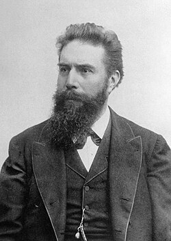
On November 8, 1895, German physics professor Wilhelm Röntgen discovered X-rays while experimenting with Lenard tubes and Crookes tubes, then began studying them intensively. He named the radiation 'X' to signify its unknown nature.
Image source: Wilhelm Röntgen
Dec 28, 1895
% complete
Röntgen wrote an initial report, 'On a new kind of ray: A preliminary communication,' and submitted it on December 28, 1895 to Würzburg's Physical-Medical Society journal, announcing his discovery to the scientific world.
1896
% complete
Röntgen's biographer Otto Glasser estimated that in 1896 alone, as many as 49 essays and 1,044 articles about the new rays were published, reflecting the enormous excitement surrounding the discovery.
Jan 1896
% complete
On reading of Röntgen's discovery, Frank Austin of Dartmouth College tested all the discharge tubes in the physics laboratory and found that only the Puluj tube produced X-rays.
Image source: Ivan Puluj
Jan 1, 1896
% complete
Along with his December submission, Röntgen sent letters on January 1, 1896 to physicians he knew around Europe, rapidly spreading news of the new rays and sparking widespread experimentation.
1896
% complete
A short time after Röntgen's landmark 1895 paper, Brandes reported that after dark adaptation and placing his eye close to an X-ray tube, he saw a faint 'blue-gray' glow which seemed to originate within the eye itself.
Feb 1896
% complete
Several weeks after Röntgen's discovery, Ivan Romanovich Tarkhanov irradiated frogs and insects with X-rays, concluding that the rays 'not only photograph, but also affect the living function.'
Image source: Ivan Tarkhanov (physiologist)
May 1896
% complete
George Albert Boulenger first mentioned Tarkhanov's work on the biological effects of X-rays in a paper delivered before the Zoological Society of London in May 1896; Gardiner's related publication with Boulenger's foreword followed in 1897.
Image source: George Albert Boulenger
1904
% complete
John Ambrose Fleming invented the thermionic diode, the first kind of vacuum tube. This idea was quickly applied to X-ray tubes, leading to heated-cathode designs.
Image source: John Ambrose Fleming
1906
% complete
Physicist Charles Barkla discovered that X-rays could be scattered by gases and that each element had a characteristic X-ray spectrum, for which he won the 1917 Nobel Prize in Physics.
Image source: Charles Barkla
1912
% complete
Max von Laue, Paul Knipping, and Walter Friedrich first observed the diffraction of X-rays by crystals in 1912, founding the field of X-ray crystallography.
Image source: X-ray crystallography
1913
% complete
Henry Moseley performed crystallography experiments with X-rays emanating from various metals and formulated Moseley's law, which relates the frequency of X-rays to the atomic number of the metal.
Image source: Henry Moseley
1920
% complete
Heated-cathode X-ray tubes, called 'Coolidge tubes', completely replaced the troublesome cold cathode or Crookes X-ray tubes — used since the 1890s — by about 1920, making X-ray generation far more reliable.
Image source: X-ray tube
1896
% complete
A child who had been shot in the head was brought to the Vanderbilt laboratory in 1896, where X-rays were used to help locate the bullet, illustrating the technology's emerging diagnostic power.
Jan 11, 1896
% complete
The first use of X-rays under clinical conditions was by John Hall-Edwards in Birmingham, England on January 11, 1896, when he radiographed a needle stuck in the hand of an associate.
Image source: John Hall-Edwards
Feb 3, 1896
% complete
Gilman Frost, professor of medicine, and his brother Edwin Frost, professor of physics, exposed the wrist of Eddie McCarthy to X-rays and collected an image of the broken bone on gelatin photographic plates.
Image source: X-ray
Feb 5, 1896
% complete
Live imaging devices were developed by Italian scientist Enrico Salvioni (his 'cryptoscope') and William Francis Magie of Princeton University (his 'Skiascope'), both using barium platinocyanide screens.
Image source: Fluoroscopy
Feb 14, 1896
% complete
On February 14, 1896, John Hall-Edwards became the first to use X-rays in a surgical operation, demonstrating their practical value in medicine just months after their discovery.
May 1896
% complete
Edison developed the first mass-produced live imaging device, his 'Vitascope', later called the fluoroscope, which became the standard for medical X-ray examinations.
Image source: Thomas Edison
1913
% complete
In 1913, a motion picture was made in Detroit showing a hard-boiled egg inside a human stomach, an early example of combining X-rays with cinematography.
Image source: Ray Park
1914
% complete
Marie Curie developed radiological cars to support soldiers injured in World War I, bringing mobile X-ray imaging to the front lines and revolutionizing battlefield medicine.
Image source: Marie Curie
1918
% complete
In 1918, X-rays were used in association with motion picture cameras to capture the human skeleton in motion, pioneering dynamic imaging of the body.
1920
% complete
In 1920, X-ray imaging was used to record the movements of the tongue and teeth in the study of languages by the Institute of Phonetics in England.
Feb 1896
% complete
Professor John Daniel and William Lofland Dudley of Vanderbilt University reported hair loss after Dudley was X-rayed, one of the earliest documented accounts of harm caused by X-ray exposure.
Image source: William Lofland Dudley
1904
% complete
Edison's glassblower Clarence Madison Dally habitually tested X-ray tubes on his own hands, developing tenacious cancer that led to amputation of both arms. He died in 1904, becoming the first known death attributed to X-ray exposure, after which Edison dropped X-ray research around 1903–1904.
Image source: Clarence Madison Dally
Aug 3, 1905
% complete
Elizabeth Fleischman, an American X-ray pioneer, died in San Francisco, California from complications resulting from her work with X-rays, another tragic casualty among early practitioners.
Image source: Elizabeth Fleischman
1908
% complete
Hall-Edwards developed cancer (then called X-ray dermatitis) sufficiently advanced by 1904 to prompt public warnings. His left arm was amputated at the elbow in 1908, four fingers on his right arm soon thereafter, and he died of cancer in 1926.
1957
% complete
Canberra proposed a ban on shoe-fitting fluoroscopes in 1957, while Switzerland prohibited the machines in 1989, reflecting growing awareness of unnecessary radiation exposure.
Image source: Shoe-fitting fluoroscope
Jul 23, 1999
% complete
The Chandra X-ray Observatory, launched on July 23, 1999, has allowed exploration of the very violent processes in the universe that produce X-rays, opening a new window on astronomy.
Image source: Chandra X-ray Observatory
2006
% complete
By 2006, medical procedures in the United States contributed much more ionizing radiation than in the early 1980s, constituting nearly half of total radiation exposure, up from 58% of human-made sources in 1987.
Image source: Medical imaging
2010
% complete
Up to 2010, five billion medical imaging examinations had been conducted worldwide, underscoring how thoroughly X-ray-based imaging had become embedded in global healthcare.
2015
% complete
As of 2015, early photon tomography (EPT), along with other techniques, is being researched as potential alternatives to X-rays for imaging applications, motivated partly by concerns over CT-related cancer risk estimated at 0.4% of current US cancers.
Image source: Tomography
Or browse the full history timeline directory, with more than 2,000 topics.
This History of X-Rays timeline was generated with the help of AI, using information found on the internet.
We work hard to keep these timelines accurate, but mistakes do get through. If you spot one, email us at [email protected] and we'll fix it for future visitors.
Generate yours with AI or build it from scratch, for free.
Export your timeline, add your own events, edit or remove AI-generated events, and much more
No credit card required.
Cancel anytime.
Cancel anytime.
You can create an unlimited number of timelines with up to 10 events on each one, customize how they look, export them to PDF, PNG, PowerPoint, CSV, and Excel, and share them with a link. Paid plans raise the event limit and add features like spreadsheet imports and collaborators.
Yes. You can cancel your subscription from your account page at any time, and you will not be charged again. Your subscription stays active for the rest of the period you already paid for.
Yes. We will email you a reminder before the annual renewal, and we will also email you a receipt.
Yes. You can email us within 15 days of any payment, and we will issue you a full refund.
Check out our pricing docs or send us an email anytime: [email protected].