-
Resources
-
Pricing
The history of histology traces how scientists developed microscopes, staining methods, and cellular theories to study the structure and function of tissues. More Less
Build a timeline like this one in minutes, for free. Start Yours →
Part of our collection of science timelines.
180
Galen's anatomical writings synthesized observations from animal dissections and experiments. His descriptions of tissues, organs, and physiological function dominated European and Islamic medical education for centuries, even though many conclusions were based on animals rather than humans.
Image source: Galen
1242
In the Sharh Tashrih al-Qanun, Ibn al-Nafis rejected Galen's proposed direct passage of blood through the interventricular septum and described circulation through the lungs. The work exemplified the continuing refinement of anatomical knowledge before microscopy.
Image source: Ibn al-Nafis
1543
Andreas Vesalius published a richly illustrated human anatomy that challenged errors inherited from Galen. Its emphasis on dissection, observation, and accurate visual representation strengthened the anatomical tradition from which histology emerged.
Image source: De Humani Corporis Fabrica Libri Septem
1590
Zacharias Janssen and his workshop are traditionally credited with producing one of the earliest compound microscopes. The attribution is debated, but early compound instruments created the technical possibility of examining structures too small for the unaided eye.
Image source: Zacharias Janssen
1624
Galileo Galilei experimented with a compound microscope, sometimes called an occhiolino, and used magnification to inspect small objects. His work helped make microscopy part of the scientific instrument culture of the seventeenth century.
Image source: Galileo Galilei
1646
Athanasius Kircher's Mundus subterraneus included observations made with early microscopes and promoted the use of magnification in natural inquiry. Some interpretations were speculative, but the book helped popularize microscopic investigation.
Image source: Athanasius Kircher
1661
Marcello Malpighi used microscopy to study organs and tissues, including lungs, kidneys, skin, and the brain. His observations of capillaries connected arteries and veins and earned him recognition as a founder of microscopic anatomy and histology.
Image source: Marcello Malpighi
1665
Robert Hooke's Micrographia presented detailed illustrations of objects viewed with a compound microscope. His description of cork compartments as cells introduced a foundational term to biology, although the structures he saw were mostly dead cell walls.
Image source: Micrographia
1672
Nehémiah Grew applied microscopy systematically to plant anatomy. His descriptions of tissues, vessels, and plant organization helped establish comparative microscopic anatomy and contributed to the later development of plant histology.
Image source: Nehemiah Grew
1674 - 1723
Antonie van Leeuwenhoek constructed powerful single-lens microscopes and observed protozoa, bacteria, spermatozoa, red blood cells, muscle fibers, and other microscopic structures. His reports greatly expanded knowledge of the living microscopic world.
Image source: Antonie van Leeuwenhoek
1757 - 1766
Albrecht von Haller's Elementa physiologiae synthesized anatomy and experimental physiology. Its treatment of tissues and organ function supported the idea that biological organization should be analyzed at progressively smaller structural levels.
Image source: Albrecht von Haller
1801
Xavier Bichat's Anatomie générale classified the human body into tissues such as mucous, serous, cellular, muscular, and nervous tissues. He emphasized tissue organization as a level distinct from whole-organ anatomy, despite rarely using a microscope.
Image source: Xavier Bichat
1824
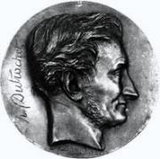
Henri Dutrochet argued that plants and animals were composed of globular elementary units and investigated osmosis across membranes. His work helped move biology toward a general theory of cellular organization.
Image source: Henri Dutrochet
1830 - 1833
In the early 1830s, Purkynĕ invented a high-precision microtome.
1831
Robert Brown identified and named the nucleus while studying orchid cells. Recognizing the nucleus as a consistent component of plant cells became an important step toward the cellular conception of living tissues.
Image source: Robert Brown (botanist, born 1773)
1838
Matthias Schleiden proposed that plants are composed of cells and that the cell is the basic structural unit of plant life. His synthesis helped provide the conceptual framework for interpreting microscopic tissue observations.
Image source: Matthias Jakob Schleiden
1839
Theodor Schwann extended cellular principles to animals and argued that cells form the fundamental units of living organisms. Together with Schleiden's work, his theory transformed histology from descriptive tissue anatomy into cellular biology.
Image source: Theodor Schwann
1841
Jakob Henle's systematic work on microscopic anatomy described tissues and organs with unprecedented organization. His studies of epithelial, connective, muscular, and renal structures influenced generations of histologists and clinicians.
Image source: Friedrich Gustav Jakob Henle
1858
Rudolf Virchow's Cellular Pathology argued that disease arises from alterations in cells and that every cell comes from a preexisting cell. This principle connected histology directly with pathology and clinical diagnosis.
Image source: Rudolf Virchow
1879 - 1882
Walter Flemming used aniline dyes to observe chromosomes and documented the stages of nuclear division, naming the process mitosis. His work established a cellular mechanism for growth, repair, and reproduction in tissues.
Image source: Walther Flemming
1891
Wilhelm von Waldeyer-Hartz popularized the term neuron and synthesized evidence that the nervous system is composed of discrete cells. The neuron doctrine became central to modern neurohistology, although its implications were initially debated.
Image source: Heinrich Wilhelm Gottfried von Waldeyer-Hartz
1900
By the early twentieth century, cytology had become a distinct microscopic discipline focused on individual cells. Its emergence allowed histologists to connect tissue architecture with cellular morphology, division, and pathological change.
Image source: Cell biology
1861
Louis Pasteur's experiments on fermentation and putrefaction provided strong evidence that microorganisms arise from existing life rather than spontaneous generation. The resulting germ theory stimulated the use of microscopy and tissue examination in infectious disease research.
Image source: Louis Pasteur
1870 - 1889
Carl Weigert and other investigators developed dyes and protocols that selectively stained bacteria and tissue components. These methods improved the microscopic diagnosis of infection and laid groundwork for specialized histochemical techniques.
Image source: Karl Weigert
1870 - 1889
Wilhelm His developed methods for embedding, sectioning, and reconstructing embryos from serial sections. His work advanced developmental histology and provided a more rigorous way to infer three-dimensional anatomy from thin slices.
1871
Charles Darwin's evolutionary synthesis encouraged comparative studies of tissues and organs across species. Histologists increasingly interpreted structural similarities and differences as evidence of common ancestry and adaptation.
Image source: The Descent of Man, and Selection in Relation to Sex
1872 - 1886
The Carl Zeiss workshop, working with Ernst Abbe and Otto Schott, produced standardized high-quality microscope objectives and optical glass. These innovations made reliable high-resolution microscopy broadly available to histologists.
Image source: Carl Zeiss
1873
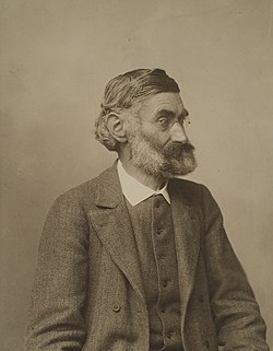
Ernst Abbe mathematically defined the limits of optical resolution and explained how numerical aperture and wavelength affect image detail. His theory guided the design of better objectives and made microscopy a more quantitative science.
Image source: Ernst Abbe
1873
Camillo Golgi introduced a silver impregnation method that selectively revealed nerve cells and their processes. Golgi staining transformed neurohistology by making complex neuronal architecture visible in black against a pale background.
Image source: Camillo Golgi
1880
Charles Louis Alphonse Laveran observed malaria parasites in the blood of infected patients. His discovery demonstrated the value of microscopic examination for identifying disease-causing organisms in clinical specimens.
Image source: Charles Louis Alphonse Laveran
1884
Hans Christian Gram introduced a differential stain that separates bacteria into Gram-positive and Gram-negative groups according to cell-envelope properties. Gram staining became a central tool in microbiology, pathology, and tissue diagnosis.
Image source: Gram stain
1886 - 1910
Albrecht Kossel characterized nucleic-acid components and nuclear proteins, including the chemical constituents of chromatin. His work connected microscopic nuclear structures with their molecular composition and helped prepare histology for cytochemistry.
Image source: Albrecht Kossel
1888 - 1906
Using modified Golgi staining, Santiago Ramón y Cajal produced detailed drawings of nervous tissue and argued that neurons are discrete structural units. His work established modern neurohistology and earned him the 1906 Nobel Prize with Camillo Golgi.
Image source: Santiago Ramón y Cajal
1903
Ultramicroscopy made it possible to detect particles below the ordinary light microscope's resolution through scattered light. It expanded the study of colloids, viruses, and fine cellular structures before the electron microscope became available.
Image source: Ultramicroscope
1870 - 1879
The combination of hematoxylin, which highlights nuclei, and eosin, which colors much of the cytoplasm and extracellular matrix, became the standard general-purpose stain of histology. H&E remains central to surgical pathology and teaching.
Image source: H&E stain
1875 - 1899
The adoption of paraffin as an embedding medium enabled tissues to be hardened, cut into thin serial sections, and mounted for staining. Paraffin processing became one of the most important practical foundations of routine histology and pathology.
Image source: Histology
1931
Ernst Ruska and Max Knoll built the first electron microscope, replacing visible light with a beam of electrons. Electron microscopy eventually revealed organelles, membranes, ribosomes, and other ultrastructures far below the resolution of light microscopy.
Image source: Electron microscope
1940 - 1959
Albert Claude combined electron microscopy with cell fractionation to investigate the internal organization of cells. His work helped establish modern cell biology and earned him the 1974 Nobel Prize with Christian de Duve and George Emil Palade.
Image source: Albert Claude
1941
Albert Coons and colleagues labeled antibodies with fluorescent dyes to identify specific antigens in tissue sections. Immunofluorescence introduced molecular specificity into microscopy and became an important tool in immunology, pathology, and cell biology.
Image source: Immunofluorescence
1950 - 1969
George Emil Palade used electron microscopy to characterize ribosomes, the endoplasmic reticulum, mitochondria, and secretory pathways. His findings connected tissue morphology to the mechanisms of protein synthesis and intracellular transport.
Image source: George Emil Palade
1953
James Watson and Francis Crick proposed the double-helical structure of DNA using model building and evidence including Rosalind Franklin's diffraction data. Molecular genetics subsequently transformed histology by explaining cellular identity and disease at the level of genes.
Image source: Molecular Structure of Nucleic Acids: A Structure for Deoxyribose Nucleic Acid
1975
Georges Köhler and César Milstein developed hybridoma technology for producing antibodies with a single specificity. Monoclonal antibodies enabled reproducible immunohistochemistry, allowing pathologists to identify cell lineages, receptors, and disease markers.
Image source: Monoclonal antibody
1980 - 1999
Advances in antibodies, antigen retrieval, enzyme labels, and automated staining made immunohistochemistry routine in diagnostic pathology. Tissue sections could now be classified by molecular markers as well as by shape and staining patterns.
Image source: Immunohistochemistry
1980 - 1989
The spread of confocal laser scanning microscopes enabled optical sectioning and three-dimensional reconstruction of fluorescently labeled specimens. Confocal methods reduced out-of-focus blur and became widely used in histology, neuroscience, and developmental biology.
Image source: Confocal microscopy
1983
Kary Mullis developed the polymerase chain reaction, making it possible to amplify selected DNA sequences rapidly. PCR brought highly sensitive genetic testing into pathology and complemented morphological examination of tissue.
Image source: Polymerase chain reaction
1990 - 2003
The Human Genome Project created a reference sequence for human DNA and accelerated the identification of genes associated with disease. Histology increasingly became integrated with genomics, transcriptomics, and molecular classification.
Image source: Human Genome Project
1990 - 2099
High-resolution scanners began converting entire glass slides into digital images that could be viewed, shared, archived, and analyzed computationally. Whole-slide imaging supported telepathology, education, research, and increasingly automated diagnosis.
Image source: Digital pathology
1994
The cloning and expression of green fluorescent protein provided a genetically encoded way to visualize cells and proteins in living specimens. Fluorescent protein technologies linked dynamic cell behavior to tissue architecture.
Image source: Green fluorescent protein
2009
High-throughput single-cell RNA sequencing made it possible to profile gene expression in individual cells rather than averaged tissue samples. The method revealed previously unrecognized cell types and states within complex tissues.
Image source: Single-cell sequencing
2015
Expansion microscopy physically enlarges a specimen through a swellable polymer while preserving molecular relationships. The technique improves effective resolution with conventional microscopes and has been applied to cells, tissues, and neural circuits.
Image source: Expansion microscopy
2017
Deep-learning systems began demonstrating strong performance in detecting and classifying structures and lesions in digitized histological slides. AI has since become an active area of research for diagnosis, prognosis, biomarker assessment, and quantitative tissue analysis.
Image source: Computational thinking
Build a timeline like this one in minutes, for free. Start Yours →
Or browse the full history timeline directory, with more than 2,500 topics.
This History of Histology timeline was generated with the help of AI, using information found on the internet.
We work hard to keep these timelines accurate, but mistakes do get through. If you spot one, email us at [email protected] and we'll fix it for future visitors.
Generate yours with AI or build it from scratch, for free.
Export your timeline, add your own events, edit or remove AI-generated events, and much more
No credit card required.
15-day money-back guarantee.
You can create an unlimited number of timelines with up to 10 events on each one, customize how they look, export them to PDF, PNG, PowerPoint, CSV, and Excel, and share them with a link. Timelines made with Timeline Maker AI keep every event the generator created, and you can add up to 5 events of your own. Paid plans remove the event limit, add spreadsheet imports and hide Preceden's branding, and the higher-priced plan also adds collaborators, a custom logo and a custom domain.
Yes. You can cancel your subscription from your account page at any time, and you will not be charged again. Your subscription stays active for the rest of the period you already paid for.
Yes. We email you a reminder 15 days before your annual renewal, and we email you a receipt after every payment.
Yes. You can email us within 15 days of any payment, and we will issue you a full refund.
Check out our pricing docs or send us an email anytime: [email protected].